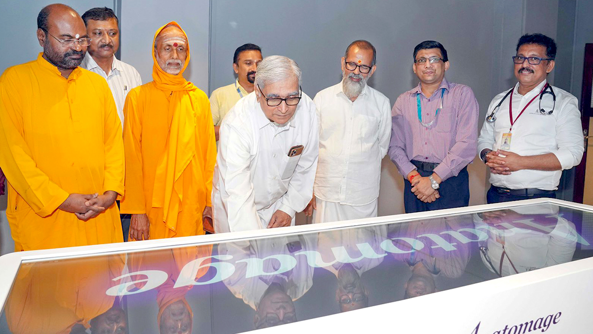
Kochi: Amrita Vishwa Vidyapeetham, Kochi, Health Sciences introduced Kerala’s first ever Virtual Anatomy Table (Anatomage Table), recently.
This introduction is a ground-breaking development for medical education, which takes teaching and learning the crucial subject of Anatomy to the next level.
What is a Virtual Anatomy Table?
The Virtual Anatomy Table is a cutting-edge, real-human-based digital platform that revolutionizes medical education by integrating advanced visualization technology with authentic cadaveric data.
It uses digitized cadavers reconstructed from frozen slices of real human bodies, providing highly detailed, life-size 3D representations.
These models include male, female, geriatric, and pregnant bodies, with anatomical precision down to 0.5 mm.
The entire vascular system is meticulously mapped, and thousands of annotated structures allow users to explore anatomy in extraordinary detail.
The Digital Anatomy Lab incorporating the “Anatomage” Table at Amrita School of Medicine was inaugurated as a special event by Dr. Prem Nair, Provost and Group Medical Director of Amrita Hospitals.
The event was attended by distinguished faculty members, including Dr. Gireesh Kumar, Principal of Amrita School of Medicine, Dr Minnie Pillay, Dr. Subramania Iyer, Dr. Asha J. Mathew, Dr. Rathi Sudhakaran, Dr. Nanditha, and other Anatomy faculty members, as well as faculty from other departments of the institute.
The Anatomage Table will augment cadaver-based learning with an immersive and interactive experience.
“Medical students can now explore and understand the intricate structures of the human body from a 3D perspective,” said Dr. Gireesh Kumar. “They can rotate, zoom, and interact with anatomical images, observe real-time clinical simulations, and study the effects of diseases on organs, gaining exceptional clarity in examining even the smallest and most complex details.”
He further went on to add, “The new facility is expected to benefit over 5,000 students at Amrita’s Kochi Health Sciences campus.”
The Anatomage Table is also an invaluable tool for diagnostics, surgical planning, and patient communication. Medical trainees can practice procedures such as dissection, craniotomy, ultrasound, and catheterization in a risk-free environment, reducing surgical errors and improving procedural accuracy.
The platform includes thousands of real-patient cases and histology scans, allowing users to study various pathologies and cellular structures.


